


Musculoskeletal Clinical Vectorial Model – Online Course
A physics-based model of muscle forces for assessing and treating mechanical spinal and joint disorders — segmental treatment of the muscles that concentrate load on the joint, systemic control of the adaptations that bring the symptom back.
The Musculoskeletal Clinical Vectorial Model:
— identifies muscle as the final effector on which origins of different nature converge — neurophysiological, biomechanical, psychosomatic — and the shortening of its parallel connective components as the mechanism that produces the symptom, in the absence of underlying disease.
Three clinical forms follow:
- nociceptive within the muscle
- nociceptive from intra-articular and discal compression
- neuropathic from radicular compression, with projection along the corresponding dermatomal and peripheral innervation territories;
— establishes what to correct and why.
Therapeutic objectives rather than protocols, leaving the therapist to decide what to do, and in what sequence, for that patient, in that session;
— acts through active exercise: isometric contractions at maximum elongation, physiological or relative, to reverse the shortening of the connective component;
— works at both levels simultaneously: segmental, on the muscles responsible for the symptom; systemic, on the redistribution of tension that would reabsorb the correction;
– quantifies the biological component of the biopsychosocial model: the peripheral mechanical substrate becomes measurable and modifiable, and with it the nociceptive and proprioceptive input sent to the central nervous system. Pain neuroscience describes how the CNS amplifies that input. The model reduces it at its source.
The model applies classical mechanics and the mechanics of materials to the musculoskeletal system:
- muscle forces represented as vectors
- the deformation of connective tissue under sustained load
- measurable and predictable force ratios between agonists and antagonists
Developed by practising physiotherapists in collaboration with a physicist and an engineer.
The physical basis of the model is formalized in a peer-reviewed article by Mauro Lastrico, indexed in Scopus, Web of Science, and EMBASE.
DOI: 10.1016/j.mehy.2026.112052.
"An Integrated Biomechanical Model Linking Connective Tissue Shortening, Segmental Load Distribution, and Vectorial Dominance to Predict Joint Degeneration Patterns"
The complete model is published in the hub article: Musculoskeletal Biomechanics — articles and publications
For Physiotherapists, osteopaths and rehabilitation professionals – CPD international
Physical Therapists and Physical Therapist Assistants · Category: General – CEU
Format 18 video modules — 32 hours on-demand · 6 hours downloadable PDF materials · Total learning time: 38 hours.
Access All video modules available on-demand for 12 months from enrolment.
Instructors Mauro Lastrico and Laura Manni, Physiotherapists
Support Direct chat with instructors for clinical questions throughout the course.
Certifications included:
38 CPD hours — The CPD Certification Service (Provider No. 21418) · For physiotherapists, osteopaths, and rehabilitation professionals.
CPD certificate issued to all participants regardless of country of residence.
45 contact hours / 4.5 CEU — Approved by the Florida Board of Physical Therapy Practice (FPTA Approval No. CE26-1318645) for Physical Therapists and Physical Therapist Assistants · Category: General · Tracked via CE Broker (Provider ID 50-54885) · Licensed professionals in other US states should verify acceptance with their own state board.
Fee €610 · Secure payment via Stripe · 2 installments available
The technical execution is teachable.
The treatment is active exercise: the skeletal segments are positioned at the angles at which the target muscles reach maximum elongation, and the patient does the work.
The real complexity lies elsewhere — in assessment, reasoning, and treatment progression, which is what the course is built around.
The reasoning has two levels.
Interpretive level
It applies the mechanics of materials to the behaviour of the connective and contractile components of muscle, and on that basis explains clinical observation: deviation and compression of skeletal structures.
The symptoms that follow can be muscular or structural.
It holds until those laws are refuted.
Applied level
It applies the same laws in reverse.
Isometric contraction at maximum available length has proved effective in re-lengthening connective tissue and restoring efficiency to the contractile components.
Verification is clinical: realignment and decompression of skeletal structures, reduction or disappearance of the symptom.
It holds until other techniques produce better results.
The model develops the work of Françoise Mézières, who through clinical observation identified muscle shortening and drew a therapeutic method from it.
The AIFIMM instructors, Mauro Lastrico and Laura Manni, trained with her in Paris between 1988 and 1990.
Their work established the physical basis of her observations and drew from it a clinical model that can be measured and verified.
This makes clinical reasoning causal, from assessment to treatment priorities: a hypothesis on the cause of the symptom, the tools to act on it, and revision of the hypothesis when the result does not follow.
Therapeutic application of the MSK clinical vectorial model ranges from the simplest cases to the most complex, including symptoms that recur or migrate.
The content is the in-person course AIFIMM has taught since 1996 — the same theoretical material, the same practical demonstrations.
A synthesis of the foundations is available in the free e-book with downloadable PDF.
Lesson 5 — Supine position work, sagittal plane. Approximately 2 hours.
A full lesson from the course, uncut: biomechanical analysis, observation criteria, practical application.
The same format as all 18 lessons of the course.
Original Italian lectures, fully dubbed in English by professional voice actors.
No registration required — watch it directly on the enrollment page.
The course starts from the foundations of the MSK clinical vector model and structured clinical assessment.
It progresses through segmental analysis — spine, lower limbs, upper limbs, TMJ — to clinical reasoning on complex cases: when the symptom is local, when it is compensatory, where to intervene first.
- Theoretical Foundations and Assessment (videos 1–4 – 8h 30min)
Causes of muscular shortening, Resistant Force and Working Force, vector analysis applied to the muscular system, three-dimensional patient assessment, manual techniques. Theory and practical demonstrations.
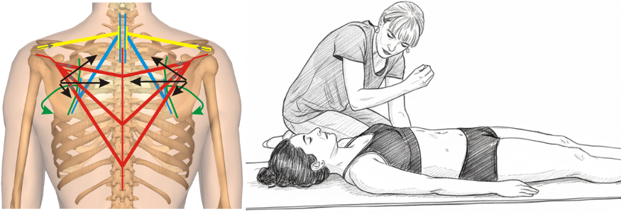
The analytical method: transforming anatomical muscles into force vectors to predict their mechanical effects. Left: Vector analysis reveals how shortened muscles create predictable force patterns affecting scapular positioning. Right: Clinical application where the therapist uses this vector understanding to guide treatment, working with therapeutic breathing to reduce resistant forces.
- Sagittal Plane Corrections (videos 5–6 – 3h 55min)
Evaluation and corrective treatment, both segmental and systemic, of the cranio-vertebro-sacral system in the sagittal plane. Theory and practical demonstrations.
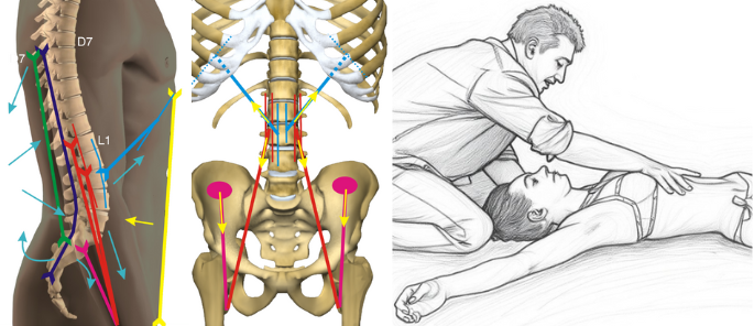
Vector analysis of forces acting between D7 and sacrum with their resultant force lines. Clinical application exemplifies how vector understanding guides therapeutic positioning - one example from the comprehensive toolkit of sagittal techniques presented in the course.
- Frontal and Rotational Plane Corrections (videos 7–9 – 5h 51min)
Evaluation and corrective treatment, both segmental and systemic, of the cranio-vertebro-sacral system in the frontal plane; evaluation and treatment of upper limb pathologies and their connection with vertebral, costal, and hyoid dysfunctions. Theory and practical demonstrations.
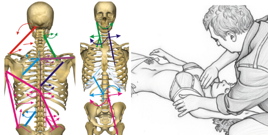
Vector analysis in the frontal plane reveals asymmetries and rotational patterns of the cranio-vertebro-pelvic system. Mechanical connections between upper limb and vertebro-costal complex guide integrated treatment - one of the specific approaches for frontal and rotational dysfunctions.
- Lower Limbs and Specific Techniques (videos 10–12 – 5h 36min)
Evaluation and treatment, analytical and systemic, of lower limb pathologies and their relationship with vertebral dysfunctions. Theory and practical demonstrations.
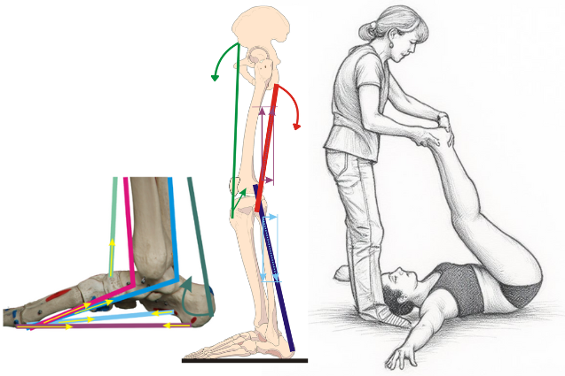
Vector representation of force lines acting on medial plantar arch and lower limb, with example of their treatment. These mechanical relationships enable both local and systemic therapeutic interventions.
- Specific Body Regions (videos 13–15 – 5h)
Distinction between primary muscular problems and those secondary to structural alterations from other systems. Evaluation and treatment of TMJ disorders and the multidisciplinary approach, dynamic analysis and identification/treatment of altered patterns, treatment of humeral and sternoclavicular subluxations. Theory and practical demonstrations.
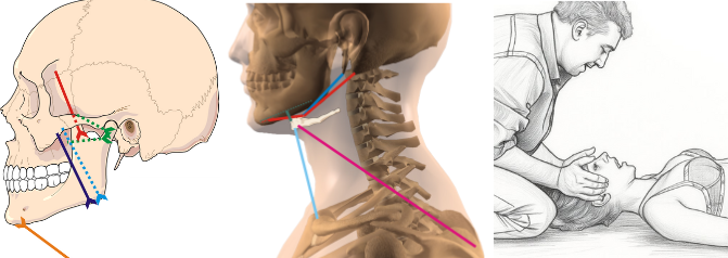
Vector analysis of temporomandibular joint, hyoid bone and cervical connections, with specific clinical application. Understanding these anatomical relationships determines whether direct treatment or multidisciplinary referral is indicated.
- Clinical Reasoning (videos 16–18 – 3h 04min)
The symptom as an expression of a local or referred problem; from static and dynamic objective examination to treatment planning. Scoliosis: evaluation and treatment. Theory and practical demonstrations.
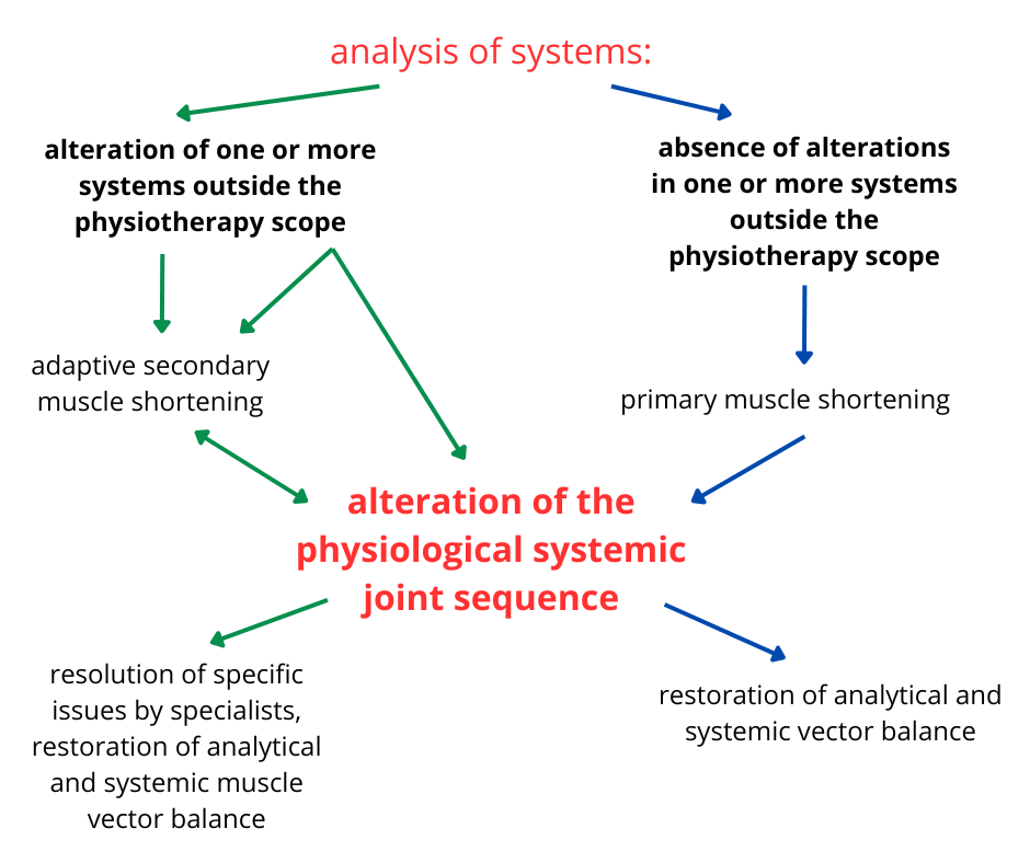
Systemic clinical reasoning: from systems analysis to treatment planning. The diagram illustrates the decision-making process for distinguishing between primary muscle shortenings (directly treatable) and secondary ones (requiring multidisciplinary approach), guiding optimal therapeutic strategy.
Each video includes theoretical lessons, live demonstrations, and downloadable PDF materials. Download full program
- 18 video modules (32 hours) — demonstrations with detailed biomechanical analysis on real patients
- Complete PDF materials organized by topic with clinical illustrations. Over 25 downloadable resources covering the theoretical foundation of the model taught in the course.
- On-demand access: available 24/7 for 12 months
- Dedicated chat with Mauro Lastrico and Laura Manni for clinical questions throughout the course
Video demonstrations include precise anatomical landmarks for positioning, patient response indicators, verification criteria, and common errors. The course provides the visual and conceptual tools to self-correct through objective patient responses: changes in joint alignment, muscle tension, and patient feedback.
Final test: 20 multiple-choice questions. Upon passing: digital certificate delivered by email.
Fee
€610 · or 2 installments of €305 (second installment due within 30 days) · Secure payment via Stripe · No hidden fees, no renewals.
Immediate access after payment.
Certifications included:
38 CPD hours — The CPD Certification Service (Provider No. 21418) · For physiotherapists, osteopaths, and rehabilitation professionals.
CPD certificate issued to all participants regardless of country of residence.
45 contact hours / 4.5 CEU — Approved by the Florida Board of Physical Therapy Practice (FPTA Approval No. CE26-1318645) for Physical Therapists and Physical Therapist Assistants · Category: General · Tracked via CE Broker (Provider ID 50-54885) · Licensed professionals in other US states should verify acceptance with their own state board.
CE Broker Tracking 20-1318645 · FPTA Approval CE26-1318645 · Effective 01/01/2026–12/31/2026. "Accreditation of this course does not necessarily imply the FPTA supports the views of the presenter or the sponsors."
The CPD Certification Service (UK) examined the biomechanical model, the teaching structure, and the coherence between stated objectives and actual content:
|
"An advanced online course providing rehabilitation professionals with a scientifically grounded model for assessing and treating musculoskeletal dysfunctions through systemic biomechanics. Based on the principles of the Mézières Method, it integrates physics, myofascial chain analysis, and vector-based muscle assessment to identify primary and secondary shortenings, optimize joint alignment, and restore functional balance. Includes 32h of video lessons and 6h of reading materials, with demonstrations, case studies, and diagnostic tools."
|

The Florida Board of Physical Therapy Practice reviewed and approved the course. From the official course record:
|
"A clinical model for the assessment and treatment of complex musculoskeletal dysfunctions, based on systemic biomechanics, vector analysis, and functional adaptation mechanisms. Participants will learn to identify residual muscular shortening, compensatory patterns, and joint compression, linking local symptoms to dysfunctions of systemic origin. The therapeutic approach includes specific manual techniques and active elongation work, aiming to restore joint function, reduce compressive forces, and reestablish mechanical balance." |
Mauro Lastrico and Laura Manni, Physiotherapists.
Over 40 years of clinical experience each.
More than 6,000 physiotherapists trained through AIFIMM, the postgraduate institute they founded in 1996.
Accredited in Italy (ECM — Ministry of Health), UK (CPD), and USA (CEU, Florida).
The MSK clinical vector model took shape across thirty years of clinical practice and shared teaching.
Teaching quality — official data
ECM is the mandatory continuing education system for healthcare professionals in Italy, managed by the Ministry of Health.
Every participant completes an evaluation, collected independently of the provider.
From 6,147 evaluations (1997–2025):
- 78% rated the training very useful
- 71% rated overall quality excellent
- 77% rated practical content delivery excellent
- Over 90% would recommend the course to a colleague
Books
Musculoskeletal Biomechanics and the Mézières Method First edition 2016 — third printing 2023 Mauro Lastrico · Demi Edizioni
Physics of the Musculoskeletal System — From the Vector Model to Clinical Reasoning Mauro Lastrico · Laura Manni · Forthcoming 2026
Articles published in indexed scientific journals
Lastrico M. An integrated biomechanical model linking connective tissue shortening, segmental load distribution, and vectorial dominance to predict joint degeneration patterns. Medical Hypotheses (Elsevier). 2026. DOI: https://doi.org/10.1016/j.mehy.2026.112052
Indexed on PubMed, Scopus, Web of Science.
Articles
The following articles have been independently evaluated and published by the CPD Certification Service (UK):
Muscle Shortening and Joint Dysfunction – Physical and Clinical Mechanisms
Body Equilibrium – A Physical-Clinical Interpretation of Human Upright Stability
Vector Analysis in Musculoskeletal Biomechanics - Part 1: Foundations and Clinical Principles
Vector Analysis of the Vertebral Column in the Frontal Plane – Part 1
Vector Analysis of the Vertebral Column in the Frontal Plane – Part 2
TMJ Biomechanical Analysis - Part 1: Systemic Relationships, Hyoid Function and Dental Influence
TMJ Biomechanical Analysis - Part 2: Muscular Mechanims, Joint Locking and Clinical Tests
Biomechanical Analysis of the Lower Limb – Part 1: Vectorial Dominances of the Hip and Knee
Biomechanical Analysis of the Lower Limb – Part 2: Vectorial Dominances of the Foot
The Muscle as Final Effector: Three Converging Systems
The biomechanical model presented in this course is not "the truth about the human body."
It is the most rigorous hypothesis we have been able to build over 40 years of clinical and theoretical work, grounded in current knowledge of physics, physiology, and biomechanics, and in the clinical observations that Françoise Mézières transmitted to us.
We know that today's knowledge will be refined and surpassed. Mézières collected the clinical observations of her time and translated them into groundbreaking insights. We translated those insights into verifiable physical and mathematical language. Others, after us, will refine, correct, and improve.
If current knowledge were complete, we would all be doing the same thing. The existence of different approaches does not mean some are right and others wrong — it means we are still exploring a complex system with limited tools.
We do not present ourselves as holders of absolute certainties. We encourage participants to test what we teach against their own clinical practice, to compare it with other frameworks, and to question what does not hold up.
Scientific rigor means building testable hypotheses, refining them, and accepting they will be surpassed.
The foundation of the AIFIMM biomechanical model rests in the international scientific literature, formalized in an article published by Elsevier, peer-reviewed, and indexed in Scopus, Web of Science, and EMBASE
DOI: 10.1016/j.mehy.2026.112052.
An Integrated Biomechanical Model Linking Connective Tissue Shortening, Segmental Load Distribution, and Vectorial Dominance to Predict Joint Degeneration Patterns — Mauro Lastrico AIFIMM instructor.
Applying the laws of physics to the musculoskeletal system, the article establishes the foundations of the model:
The connective component of muscle shortens.
The shortening is proportional to the force and duration of contraction.
The muscular system has a dual role: joint stability and movement.
Shortening alters the muscle's force, which divides into two components: Resistant Force, which the muscle opposes to its own lengthening, and Working Force, available for movement.
The two are inversely proportional.
Clinically, a shortened muscle is a shortened cord: at rest, it already holds the vertebral and articular axes deviated and under compression, producing the symptoms of mechanical conflict.
→ connective tissue mechanics
The role of antigravity muscles.
In maintaining balance, the muscular system does not work against gravity: it keeps the individual centres of gravity — and the overall centre of the body's weight — on the same vertical as the ground reaction force R.
When the centres are aligned, small variations in baseline tone suffice to compensate the body's automatic movements.
When they are misaligned, baseline tone must rise, producing residual shortening that misaligns the centres further: a self-reinforcing cycle.
Clinically, the common cause of misalignment is the shortening of the connective components.
→ body balance and ground reaction force
Vector analysis.
Every muscle, beyond its anatomical reality, can be represented as a line of force defined by its points of insertion on the bones.
Vector decomposition applies to these lines of force.
Muscles are not arranged symmetrically around joints: by number, length, and obliquity, some prevail over others.
Clinically, this makes it possible to identify these dominances between agonists and antagonists and to predict the direction of misalignment.
→ vector analysis of muscle forces · individual body regions
The same physical laws that produce shortening govern its reverse: lengthening.
The technique of choice is isometric contraction at maximum elongation, physiological or relative.
With the insertions fixed, the contractile elements — actin and myosin — contract actively and pull on the connective components arranged in parallel.
Because connective tissue has a lower elasticity coefficient than the contractile elements, the muscle gains length at the end of the contraction.
Clinically, joints and vertebral segments move back toward their physiological sequence, reducing intra-articular, discal, and radicular compression.
A lengthened muscle also lowers its Resistant Force in favour of Working Force, improving contractile capacity in movement.
→ active exercise
Structure and function
Shortened muscles shift the skeletal axes toward the dominant vectors.
In movement, the antagonist must then produce the movement and, in addition, overcome the Resistant Force of the shortened dominant — as in the relationship between the internal and external rotators of the humerus, where the former prevail.
Clinically, if the structure is altered, so is the dynamics: correcting the skeletal axis improves the movement in turn.
Muscles do not shorten in isolation. Every muscle shares insertions, force lines, and mechanical relationships with others. When one muscle shortens, the forces on its joint change, and the system adapts to hold its balance.
That adaptation is the misalignment of the body's centres of gravity: to keep the overall centre over the base of support, the segmental centres shift out of line. The cost is loss of physiological alignment in other segments — sometimes far from the original shortening.
Shortening alters the axes; altered axes require compensatory activation; sustained compensation produces further shortening. This self-reinforcing cycle raises Resistant Force at the expense of Working Force, moving the system away from the state of maximum adaptability — where Working Force dominates, FL ≫ FR — toward rigidity.
Clinically, this is the physical reason symptoms recur and migrate.
The misaligned centres of gravity still demand a higher baseline tone to hold the erect station; that raised tone re-shortens the muscle.
The cycle that produced the alteration is still running. So when a correction is applied only to the symptomatic segment, the systemic forces that generated it are still active: the system reabsorbs the correction, or keeps it at the expense of another region.
Recurrence and migrating symptoms are not treatment failures — they are the clinical sign that the cause lies beyond the treated segment.
This is why the symptomatic joint may not be the most compromised: it can simply be the point where the system's adaptive capacity was first exceeded.
Distinguishing a local problem from a systemic one is the task of clinical reasoning.
Two levels, simultaneously.
When the problem is systemic, treatment operates on two levels at once: segmental, on the muscle group responsible for the symptom; systemic, to stabilise the result over time.
The segmental work identifies the shortened muscles behind the vector dominances and intervenes on them directly.
The systemic control monitors how tension redistributes during treatment, so the local correction is not absorbed elsewhere.
Clinically, segmental work modifies the system; systemic control lets the system hold the modification. Integrating the two levels is clinical reasoning.
Tracing the symptom back to its cause makes it possible to set therapeutic priorities: what to treat, in what order, and how to verify the effect.
The result is a methodology tailored to each patient, not reducible to a protocol.
A musculoskeletal symptom can be local, referred, or systemic.
This distinction determines the therapeutic strategy and whether the result will hold over time.
Beyond history-taking and imaging, assessment draws on specific tools:
— the static examination, local and systemic, identifies the most deviated vertebral axes and joints; the magnitude of the deviation indicates whether a joint is the source of the symptom or a physiological adaptation below threshold;
— the dynamic examination identifies altered movement patterns, distinguishing a symptom sustained by movement disorganisation from one driven by structural mechanical conflict;
— dermatomal and peripheral innervation maps identify referred symptoms, where the symptomatic region and the cause do not coincide;
— tests on other structures identify when the shortening is maintained by a disturbance outside physiotherapeutic competence: its removal, through interdisciplinary collaboration, is what makes the treatment result stable.
The model is measurable, subjectively and objectively. Treatment must produce stable, progressive change on three levels:
— the symptom;
— the vertebral and articular axes, identified through the muscular vector dominances that sustain the symptom;
— the alignment of the body's centres of gravity on the ground reaction force.
If the symptoms do not regress, recur, or migrate, the initial hypothesis is revised.
→ clinical reasoning
The biomechanical model does not replace neurophysiological approaches to pain.
It addresses a specific and complementary dimension: the mechanical conditions that can initiate, maintain, or amplify the nociceptive input over time.
Central sensitisation, altered pain processing, and psychosocial factors are recognised contributors to chronic pain.
But a persistently compressed disc, a joint maintained in a non-physiological axis by shortened dominant vectors, or a mechanical conflict sustained by connective resistance are also measurable realities — and they can be modified.
The model works on the peripheral mechanical substrate.
When that substrate is contributing to the clinical picture, reducing compressive forces and restoring vectorial balance can change the mechanical input that the nervous system is processing.
This does not contradict pain neuroscience — it addresses the dimension that pain neuroscience does not treat directly.
The two frameworks are not competing explanations. They operate on different levels of the same problem.
Why muscles shorten and what happens to joint biomechanics
A consistent phenomenon is observed in clinical practice: even in the absence of specific pathologies, muscles tend to progressively shorten over time, altering both static alignment and joint dynamics.
The model explains this phenomenon through the physical laws of material deformation.
From a biomechanical perspective, muscle is not a homogeneous structure: the contractile component behaves as a reversible elastic material, while the connective component exhibits plastic behavior, retaining residual deformation proportional to the product of force × time.
This is not pathology — it is physics applied to biological tissues.
Since muscle always acts as a compressive force and the skeleton passively adapts to force resultants, muscular shortenings become the primary determinant of joint axis alterations.
On these foundations rests the RF–WF model, the core of the biomechanical interpretation:
A shortened muscle is simultaneously "too strong" from a static perspective (high Resistant Force, altering joint alignment) and inefficient from a dynamic perspective (reduced Working Force and increased compensations).
Resistant Force and Working Force are inversely proportional: as one increases, the other decreases.
This paradox explains why muscle strengthening, in the presence of structural shortenings, often fails to resolve the problem — and can worsen it.
Only by reducing Resistant Force can mechanical efficiency and function be restored.
When you palpate a shortened muscle, you are not just feeling "tension" — you are perceiving the coexistence of two problems: tissue that blocks the joint (high RF) and that simultaneously cannot do its job (low WF).
It is like driving with the handbrake engaged: the harder you press the accelerator, the more force you need to move, but that force does not move the car — it fights the brake.
This is why asking a patient to "strengthen" that area often worsens pain: you are adding power to the engine without releasing the brake.
Why joint alterations follow specific and predictable directions
Once it is clear why muscles shorten, the next question is: why do joint alterations follow recurrent and predictable directions?
This is made possible by vector analysis of muscular forces.
Muscles are not distributed symmetrically around joints.
Within every agonist–antagonist system, intrinsic anatomical asymmetries exist — in muscle number, force line length, and angle of application — creating genuine vector dominances, independent of training or voluntary control.
When Resistant Force increases, these dominances manifest first and drive the loss of physiological joint sequencing.
These are not "random compensations" — they are the predictable outcome of physical laws applied to anatomy.
This is why certain alterations are clinically recurrent. Some examples:
- In the glenohumeral joint, the humerus tends toward internal rotation and anterior displacement
- The scapulae tend toward adduction with reduction of physiological thoracic kyphosis
- The foot tends toward supination and cavus, often compensated proximally
In clinical practice, when you observe these patterns on the patient's body, the humerus is already internally rotated before you ask for any movement. It does not "internally rotate" — it is already there.
Like a crack in a wall that follows lines of least structural resistance. The anatomical dominance is already active in static posture, and movement merely amplifies it. This is why assessment begins with observation: it already tells you where to look.
Clinical reasoning changes fundamentally: you start from knowledge of anatomical dominances, predict the alterations, verify them during assessment, and rapidly identify the responsible muscular vectors.
This predictive capacity is confirmed in neurological conditions as well: when central inhibitory control is lost, as in spastic hemiparesis, the same anatomical dominances emerge in amplified form. Different mechanisms, same structural reality.
The connection with the RF–WF model is direct: when Resistant Force increases, subdominant vectors can no longer compensate, and the joint deviates along anatomically dominant directions.
In this model, muscle shortening is the final outcome of tone regulation processes involving multiple systems.
Regardless of the initial trigger, muscle is the final effector — the structure through which the body realizes adaptation. The model distinguishes different levels of origin.
Neurophysiological level: basal tone is regulated by sensory–motor integration circuits.
The nervous system uses muscle tone as a protective strategy, through pain reflexes that may activate after pain onset (reactive response) or in anticipation of it (preventive response).
When these contractions are maintained over time, they involve the connective component and transform into structural shortenings. This level lies fully within the scope of physiotherapy, because it manifests in the musculoskeletal system.
Biomechanical level: increased tone represents an adaptive response to altered loads, joint axes, or centers of gravity.
The system preserves equilibrium at the cost of a self-reinforcing cycle: mechanical alteration → increased tone → shortening → further alteration.
The result is increased Resistant Force, reduced Working Force, loss of efficiency, and progressive rigidity. This level also belongs to physiotherapy, because it produces observable and treatable joint alterations.
There is also a psychosomatic level, documented in the literature, in which prolonged emotional states modulate muscle tone through neurovegetative and central pathways, potentially leading to structured orthopedic presentations.
Whatever the level of origin, the common denominator remains the same: muscle is the final effector.
Very different clinical histories may therefore converge toward similar shortenings and compensatory patterns.
In practice, the shortened muscle is often the "final stop" of very different pathways.
One patient with chronic low back pain may present structural shortenings originating from altered mechanical loading; another from protective pain strategies maintained for years after trauma; another from distant compensations — an unresolved foot problem that modified weight-bearing patterns.
The final pattern — accentuated lordosis, anterior pelvic tilt, shortened psoas — may appear identical.
This is why assessment does not stop at reading "what you see," but seeks to understand which level is actively maintaining the problem, to avoid treating only the final effect.
The task of the model is not to explain every possible cause, but to interpret what is biomechanically relevant: distinguishing between primary cause, muscular adaptation, and articular consequences.
Why systemic analysis is a clinical necessity
In 1947, Françoise Mézières formulated what she called her capital observation: the numerous muscles behave as a single muscle, too strong and too short.
Today this insight finds explanation in complex systems theory: systems in which many elements interact interdependently.
The human musculoskeletal system is, in every sense, a complex system. This means that no region functions in isolation and that any local intervention inevitably produces adaptations at a systemic level.
The concept of muscle chains represented an initial step beyond segmental thinking. This model takes it further, placing it within a biomechanical framework grounded in physical laws and force analysis.
In a complex system, the symptom does not necessarily coincide with the site of the problem.
Local pain may be the expression of an altered systemic organization, and attempts at isolated correction can be not only ineffective, but mechanically counterproductive.
This is why instructions like "stand up straight" or many voluntary self-corrections are often ineffective: they increase overall tone and Resistant Force, worsening the system's energy balance.
Another characteristic of complex systems is the presence of emergent abilities.
When specific muscles become ineffective due to excessive shortening, the system does not stop: it bypasses them, activates alternative synergies, and develops substitution patterns.
This is clearly visible during movement: you ask the patient to raise the arm and the first thing that elevates is the shoulder, or you see the pelvis shift when only the femur should flex.
This is not "incorrect" — it is the only strategy the system has found to complete the requested movement when the muscles that should perform it are too shortened to work.
It is like taking a winding alternative road when the direct route is blocked: it works, but it costs far more in energy and wear.
This explains why isolated strengthening of subdominant muscles does not resolve the problem, and why certain monoarticular muscles are systematically excluded from movement.
In this model, a system functions efficiently when it operates at the edge of chaos — that zone of maximum flexibility and adaptability where the body is ready to respond to any stimulus without rigidity — a condition in which Working Force prevails over Resistant Force.
When Resistant Force increases, the system becomes rigid, energy expenditure rises, and physiological joint sequencing is lost.
An efficient system moves fluidly, with no visible effort, with smooth transitions between segments. When RF increases, movement becomes fragmented, "jerky," with visible pauses and compensations.
The patient tells you "I struggle with simple things" — not because of weakness, but because every gesture requires three times the energy.
It is like pushing a shopping cart with the wheels locked: you push hard but barely move, and you tire immediately.
From symptom to the distinction between local dysfunction and systemic organization
In musculoskeletal pain, the symptom always represents real clinical information, but it does not automatically coincide with the cause of the problem. In some cases the pain is genuinely local; in others, the symptomatic joint represents the outcome of an adaptive organization involving multiple regions.
Clinical reasoning is founded on the need to distinguish between local and referred pain.
The muscle tone observed in clinical practice is the result of neurophysiological and biomechanical regulation processes aimed at maintaining stability and continuity of movement.
When these adaptive strategies persist over time, they may involve the connective component of muscle, transform into structural shortenings, and alter physiological joint sequencing.
Pain often emerges when the system's adaptive margins are reduced.
In the model, clinical reasoning integrates:
- observation of static and dynamic patterns
- analysis of vector dominances
- differentiation tests aimed at determining whether the symptomatic region is the primary cause or the site of expression of a broader adaptive organization
When pain originates locally, intervention on the symptomatic region can be resolutive.
When pain is referred, treating exclusively the site of the symptom produces transient improvements that will not persist over time.
In practice, it is like a warning light on a car dashboard: it could be a faulty sensor (local problem — replace the sensor and you are done) or an overheating engine (systemic problem — the light is just the message).
In the first case, you treat where it hurts and the patient improves stably.
In the second, the shoulder pain you treat may calm down for a few days, but if the problem originates from a blocked pelvis forcing the spine to compensate, the shoulder will hurt again until you address the primary restriction.
This is why differentiation tests are fundamental: not to "prove" that pain is referred, but to avoid chasing the symptom without modifying the forces that regenerate it.
A key step in clinical reasoning is the distinction between primary and secondary muscle shortenings.
In primary shortenings, the muscular system is the origin of joint misalignment, and vector rebalancing can be stable and effective. In secondary shortenings, the muscle represents an adaptation to dysfunction originating from other systems, requiring multidisciplinary assessment to avoid recurrence.
This is why some improvements last and others do not. Not because treatment "works or doesn't work," but because the forces responsible for the misalignment have, or have not, been correctly identified and modified.
The following simplified case illustrates how clinical reasoning is applied within the model. Real clinical situations are often more complex and require multiplanar assessment and continuous adaptation, but the decision-making logic remains the same.
Clinical case A patient presents with anterior shoulder pain and limited abduction. No recent trauma.
Phase 1 — Prediction through vector analysis Based on anatomical dominances, vector analysis predicts internal rotator dominance. When Resistant Force increases in these muscles, the humeral head tends to shift anteriorly, reducing the subacromial space.
Phase 2 — Verification during assessment Physical examination confirms the predicted pattern:
- anteriorized humeral head
- static internal rotation of the humerus
- scapular adduction
Phase 3 — Decision sequence Intervention priorities are defined by force analysis, not by predefined protocols:
- address the subscapularis first (dominant vector in anteriorization)
- verify scapular repositioning, as adductors may maintain residual tension
- assess cervical compensations, frequently associated with scapular rigidity
Each step is guided by mechanical reasoning and continuously adjusted according to observed responses.
Phase 4 — Immediate verification At the end of the session:
- has the humeral head repositioned?
- has abduction improved?
- is the system more elastic, or have new rigidities emerged?
If yes, the causal vector has likely been correctly identified. If not, reassessment is required: are there unrecognized compensations? Is the shortening primary or secondary?
This is the clinical reasoning taught in the course: not "what to do for the shoulder," but how to analyze the forces altering the shoulder and adapt intervention based on system response.
In real clinical practice, every patient requires individual assessment, consideration of systemic compensations, and strategies that evolve continuously in response to mechanical feedback.
How the model guides clinical intervention
The objective of treatment is to modify the forces maintaining the system in a mechanically inefficient state, reducing Resistant Force (RF) and restoring genuinely available Working Force (WF).
This requires a fundamental distinction: the two components of muscle tissue — contractile and connective — respond to different stimuli and cannot be treated with the same approach.
Spontaneous movement, though essential for function, is insufficient to re-lengthen a structurally shortened muscular system: it always respects the limits already in place and the boundaries the nervous system has accepted as safe.
It is like trying to touch your toes: you reach as far as your body "allows" you to go, even if you push. That limit is not structural — it is the point where the nervous system says "stop, further is dangerous." Spontaneous movement always respects this safety boundary. The shortened connective tissue lies beyond that boundary: reaching it requires guided work, where the therapist progressively brings the patient beyond the habitual limit, under controlled and safe conditions.
When shortening involves the connective component, length recovery requires guided therapeutic intervention capable of bringing tissue beyond the limits of spontaneous adaptation.
Acting only on muscle tone, mobilizing or passively stretching, may be effective on the contractile component but remains insufficient on the connective substrate responsible for residual shortening.
This is why the model employs isometric contractions performed in maximum physiological or relative elongation, within a strategy continuously adapted to system response.
The technique is never applied automatically. The same maneuver can produce opposite effects depending on the mechanical context: if performed below the correct threshold, it can further increase Resistant Force. This is why precision of positioning, reading of dominances, and continuous observation of patient response are clinically decisive.
Treatment always follows a dual logic: resolve the specific mechanical conflict generating the symptom, and verify that the local correction does not generate compensations or joint misalignments elsewhere.
An intervention that improves the local area but rigidifies the system is destined to fail. A purely "global" approach that does not address the real conflict may improve the general sensation without solving the problem.
In practice, you see patients who leave the session feeling "looser," breathing better, feeling freer — but if you ask them to repeat the movement that caused pain, the conflict is still there. It is like loosening every bolt on a machine except the one that is stuck: everything feels softer, but the part does not move. You need to find where the tension truly concentrates — the shoulder that does not abduct, the pelvis that does not rotate, the knee that does not extend — and work there with precision, without rigidifying everything else.
Every observable configuration, even when it appears pathological, represents the best adaptive solution the system has found at that moment. Improvement is not defined by visual symmetrization, but by the reduction of overall tension, increased systemic space, and recovery of functional efficiency.
Treatment effectiveness criteria are not limited to immediate symptom relief. At the end of a session, three conditions must be simultaneously present: local improvement, absence of new compensatory strategies, and greater system adaptability. If even one is missing, the result tends to be unstable.
When you verify at the end of a session, you do not just ask "has the pain gone?" but also "how does the patient move now?" and "where has the tension gone?"
If the shoulder has unblocked but the patient is now holding the neck rigid, you have moved the problem. If the patient feels better but movement is still fragmented, you have reduced the symptom but not the inefficiency.
When all three criteria are present together — reduced pain, fluid movement, more elastic system — the improvement is far more likely to persist. When even one is missing, the result risks being transient.
This is why the model does not propose standardized sequences but an adaptive clinical strategy. Intervention priority varies according to the dominances present and system response: sometimes it is necessary to work on systemic aspects first, sometimes to address the segmental conflict directly, always avoiding overall rigidity.
In clinical application, this approach proves particularly effective in chronic, recurrent, or resistant cases — when symptoms reappear despite previous treatments, often because the forces regenerating them have not been identified and modified.
Treatment is always individual, active, and guided. It requires time, listening, and observation, because the clinician's task is not only to intervene, but to understand and explain.
Vector analysis thus becomes both a clinical and a communicative tool: it allows the construction of verifiable reasoning and makes the patient an active participant in the therapeutic process. This shared understanding is one of the main factors explaining the stability of results over time.
The biomechanical model rests on three complementary forms of validation.
Peer-reviewed theoretical publication — The theoretical foundations of the model are published in Medical Hypotheses (Elsevier), indexed in Scopus, Web of Science, and EMBASE. The paper formalizes, in terms of vector mechanics, the cumulative shortening of connective tissue, segmental load redistribution, and the vectorial dominance of muscle forces, generating specific and falsifiable predictions about joint degeneration patterns.
Read the article: https://doi.org/10.1016/j.mehy.2026.112052
Mechanistic reasoning — The interpretive level is grounded in physical laws applied to the musculoskeletal system: vector mechanics, the behavior of connective tissue under sustained load, and the measurable relationships linking muscular shortening, the Resistant Force / Working Force ratio, and articular conflict. These principles are internally consistent, independently verifiable, and open to refutation by physical and mathematical scrutiny.
Evidence-informed clinical observation — Over 40 years of systematic clinical work have produced consistent, measurable outcomes: changes in joint alignment, symptom reduction, and — most tellingly — coherence between the forces predicted by vector analysis and the axial changes observed after treatment. Where prediction and outcome diverge, the working hypothesis is revised.
The therapeutic technique — The model works through isometric contractions at maximum physiological elongation. Its core parameter — the length at which the muscle contracts — is supported by evidence generated entirely outside this model.
A systematic review in the Scandinavian Journal of Medicine & Science in Sports, examining isometric training specifically, found that contractions performed at longer muscle lengths produce hypertrophy at 0.86–1.69% per week against 0.08–0.83% at shorter lengths, together with greater transfer to dynamic performance. Tendon structure and stiffness respond to the same parameter, provided contraction intensity exceeds 70%.
Two further systematic reviews published in 2025 point in the same direction for longitudinal adaptation, with mixed evidence on the underlying structural mechanism: training at longer muscle lengths produces greater increases in muscle size and fascicle length than training at shorter lengths.
This body of work measures hypertrophy, strength, and tendon adaptation in healthy and athletic populations. It does not measure the clinical outcomes of this model. What it establishes is that the mechanical parameter the model is built on — contract the muscle at its maximum available length — is the parameter that produces structural change in length. That is what the model asks of it.
External evidence on the source technique — The therapeutic tools used in this model derive from the technique Françoise Mézières developed. Five randomised controlled trials on that technique have been published in indexed journals, on populations ranging from elite rhythmic gymnasts and professional footballers with low back pain to patients with Parkinson's disease. Measured outcomes include pain intensity, spinal mobility, functional capacity, and dynamic performance; one trial is double-blind and uses instrumented spinal measurement.
A separate systematic review and meta-analysis of seven trials, covering 334 patients, examined Global Postural Re-education — the approach Philippe Souchard derived from Mézières — and found reductions in pain and improvements in function against no treatment.
Two limits define what this evidence does and does not do. It concerns the source technique, not the AIFIMM clinical vector model. And while the meta-analytic comparison against no treatment is favourable, comparison against alternative active therapies remains inconclusive, given heterogeneity and risk of bias across the included trials.
What the trials establish is that the therapeutic tool has been tested by independent groups, under controlled conditions, with measurable results. What the model adds is the reasoning that decides where to apply it.
Why large-scale trials on the specific model are limited — Each treatment requires individual biomechanical assessment, continuous adaptation of strategy, extended one-to-one sessions, and therapists with advanced specific training. This individualization is central to the approach — and it is exactly what makes it hard to standardize for a large randomized trial. The difficulty is not specific to this model: individualized manual and rehabilitative interventions face the same problem across physiotherapy, which is why the field increasingly relies on pragmatic trials and single-case (N-of-1) designs alongside classical RCTs. Accordingly, the model is supported not by a single large trial but by the convergence described above: mechanistic reasoning grounded in physics, consistent clinical outcomes, and coherence with the external evidence on the techniques it employs.
What is vector analysis in musculoskeletal biomechanics? Vector analysis represents each muscle as a line of force defined by magnitude, direction, and point of application. When multiple muscles act on the same joint, their forces combine into a resultant that determines joint positioning. This course teaches how to use vector analysis to predict joint alterations, identify causal muscles, and guide treatment decisions.
What is the difference between Resistant Force and Working Force? A structurally shortened muscle generates two simultaneous forces: Resistant Force (RF) — permanent traction on bone insertions that alters joint alignment — and Working Force (WF) — the muscle's actual capacity to produce useful movement. RF and WF are inversely proportional: as one increases, the other decreases. This relationship explains why strengthening a shortened muscle often worsens symptoms.
How does this approach differ from standard biomechanics courses? Most biomechanics courses focus on assessment and analysis. This course connects vector-based assessment directly to therapeutic decision-making: which muscles to treat, in what sequence, and how to verify the result. The model is predictive — anatomical dominances allow you to anticipate joint alterations before examining the patient.
What is the Mézières technique and why is it used in this course? The Mézières technique uses isometric contractions performed in maximum physiological elongation to reduce structural shortening of the connective component of muscle tissue. It was selected for this course because of its mechanical coherence with the vector-based biomechanical model. The interpretive framework is universal; the technique adopted is Mézières.
Can this model be integrated with other therapeutic approaches? Yes. The interpretive framework — vector analysis, RF-WF mechanics, anatomical dominances — is independent of the therapeutic tool used. It provides clinical reasoning criteria that can inform any manual or exercise-based approach, provided mechanical coherence is respected.
What conditions can be treated with this approach? The model applies to orthopedic joint and spinal pathologies, chronic musculoskeletal symptoms unresponsive to standard treatments, pain that migrates between body regions, recurrent issues without clear traumatic cause, and persistent functional limitations post-surgery.
Is the course delivered in English? Yes. All video lectures are original Italian recordings fully dubbed in English by professional voice actors. All written materials and resources are in English. You can verify the audio quality by watching the free 2-hour sample lesson.
Are CPD/CEU credits recognized in my country? UK: 38 CPD hours, fully recognized. USA (Florida): 45 contact hours / 4.5 CEU, officially approved. EU, Australia, New Zealand: CPD is widely recognized. Canada: often recognized — check with your provincial regulatory body.
Do I need advanced physics or mathematics? No. Biomechanical principles are introduced progressively through clinical examples. If you understand basic anatomy and physiology, you have the foundation needed.
How much time do I need per week? The course is entirely self-paced with 24/7 access for 12 months. Most participants complete it in 3–6 months, dedicating 2–4 hours per week.
Is there instructor support? Yes. Dedicated chat with Mauro Lastrico and Laura Manni for clinical questions, case discussions, and clarifications throughout the 12-month access period. Response time typically within 24–48 hours.
Can I download the videos? Videos are streamed on-demand (not downloadable) and available 24/7 for 12 months. All PDF materials are downloadable.